
结膜良性增生性疾病的光学相干断层扫描特征
[赵丰平 , 杜持新, 蔡思捷]
, 杜持新, 蔡思捷]
|
|


第一作者:赵丰平(ORCID:0000-0003-2415-2968),Email:375636236@qq.com
目的 应用眼前节光学相干断层扫描(AS-OCT)对睑裂斑和翼状胬肉进行结构成像和厚度测量,以了解病变的结构特征。方法 病例对照研究。利用Optovue光学相干断层扫描血管造影仪(OCTA)的前节模块对2018年1-4月就诊于浙江大学医学院附属第四医院眼科的25例(30眼)研究对象进行鼻侧球结膜成像,根据诊断分成睑裂斑组8例(10眼)、翼状胬肉组9例(10眼)和正常眼组8例(10眼)。分层测量距离巩膜突0.0、0.5、1.0、1.5、2.0、2.5 mm 6个位点结膜上皮层和固有层组织的厚度。组内各位点比较采用重复测量方差分析,组间比较采用单因素方差分析。结果 与正常眼组和睑裂斑组相比,翼状胬肉组在0.5 mm处结膜上皮层明显增厚( P<0.05),其余位点差异无统计学意义。在鼻侧2.5 mm范围内,与正常眼组相比,翼状胬肉组和睑裂斑组的固有层均增厚( P<0.05),尤以胬肉组织增厚程度明显( P<0.05)。结论 AS-OCT可清晰成像结膜良性增生性疾病眼的结膜组织结构,并可进行较为精准的定量诊断和评估。翼状胬肉和睑裂斑以固有层增厚为主。
Objective: To use anterior segment optical coherence tomography (AS-OCT) for structural imaging and accurate measurements of pinguecula and pterygium in order to understand the structural features of these lesions.Methods: This study was a prospective parallel control study. Lateral bulbar conjunctiva of 25 cases (30 eyes) that included pinguecula (10 eyes of 8 cases), pterygium (10 eyes of 9 cases), and normal eyes (10 eyes of 8 cases), were enrolled from the Department of Ophthalmology of the Fourth Affiliated Hospital of Zhejiang Medical University from January to April 2018. The anterior segment module of an Optovue optical coherence tomography angiography (OCTA) was used for measurements. The thickness of the bulbar conjunctiva epithelium and propria tissues at sites at a distance of 0.0 mm, 0.5 mm, 1.0 mm, 1.5 mm, 2.0 mm and 2.5 mm from the scleral process were stratified within the 2.5 mm range, were measured, compared and further analyzed. The data were described by means of mean ± standard deviation. Repeated measurement analysis of variance was used for comparison of each point in the group, one-way analysis of variance was used for comparison between groups.Results: Compared with normal eyes and pinguecula eyes, pterygium showed thickened epithelium in 0.5 mm point ( P<0.05). In the 2.5 mm range on the nasal side, pterygium and pinguecula showed thickened subepithelium tissues ( P<0.05), especially for pterygium, compared to normal eye tissue.Conclusions: AS-OCT can image microstructures in benign conjunctival proliferative diseases, resulting in accurate quantitative diagnosis and evaluation. The thickness of subepithelium tissues increases in pinguecula and pterygium.
光学相干断层扫描(OCT)是一种新型横断面成像技术, 具备高分辨率、非侵袭性、可重复性高等优势, 可用于眼科影像学诊断[1]。近年来, 前节OCT(Anterior segment OCT, AS-OCT)模块拓展了该技术的应用领域, 可获得角膜厚度、虹膜厚度等准确的眼前节数据[2, 3, 4], 广泛应用于各种眼前节疾病的诊断和定量分析[5, 6, 7]。
Kieval等[8]利用前节OCT进行了球结膜增生性疾病的结构成像, 发现OCT能很直观地鉴别睑裂斑和翼状胬肉及结膜上皮肿瘤, 但是未进行系统的精确测量。
本研究拟采用高分辨率的眼前节OCT技术对睑裂斑、翼状胬肉的各部分结膜上皮和固有层的厚度进行测量, 并与正常眼进行对比分析, 以探讨结膜良性增生性疾病的超微结构特征和差异性, 并提供精准的量化数据。
正常眼入组标准为眼表不患有任何结膜增生性疾病患者。翼状胬肉入组标准为静止期的且头部前端与瞳孔中心距离≥ 5 mm的轻度患者[9], 翼状胬肉和睑裂斑患者排除患有影响研究结果的其余眼部情况(如球结膜水肿、色素痣等肿物等), 排除外伤史、眼球手术史、眼球震颤、无法固视等无法配合检查获取有效图像者。所有患者记录性别、年龄, 同时行裂隙灯显微镜(日本Topcon公司)照相记录病变位置。
选取2018年1-4月浙江大学医学院附属第四医院眼科就诊的25例(30眼)正常眼和翼状胬肉、睑裂斑病例。本研究遵循赫尔辛基宣言原则, 并获得浙江大学附属第一医院伦理委员会批准(2018科研快审第749号)。所有入选患者均了解本研究的目的和内容, 并签署知情同意书。
所有入选患者使用美国Optovue的光学相干断层扫描血管造影仪(Angio OCT, OCTA, 美国 Optovue公司)的前节模块进行眼前段成像。以840 nm波长的激光作为相干光源采集眼前段图像(角膜前节轴向分辨率≤ 5 μ m, 组织横向分辨率≤ 15 μ m, 扫描速度≥ 70 000次A扫描), 检查时患者取坐姿, 注视外置光标, 于外眦3点或9点位, 选用标准眼前节扫描模式, 通过操作手柄移动仪器的探头, 调节焦距及扫描位置, 对焦, 使监视器图像最清晰, 完成测量。扫描线水平宽度8 mm, 对检查眼鼻侧3点或9点位各进行3次图像采集, 选取信号值最高且最清晰的图像予以保存进行分析。均由同一位操作熟练的技师完成。
图像收集后, 用设备自带的标尺工具测量距离巩膜突(图像中表现为巩膜向内隆起)[9]0.0、0.5、1.0、1.5、2.0、2.5 mm的6个位点结膜上皮和结膜固有层的组织厚度(见图1)。读片和测量时均手工校准巩膜突位置, 每个参数均测量3次取平均值。
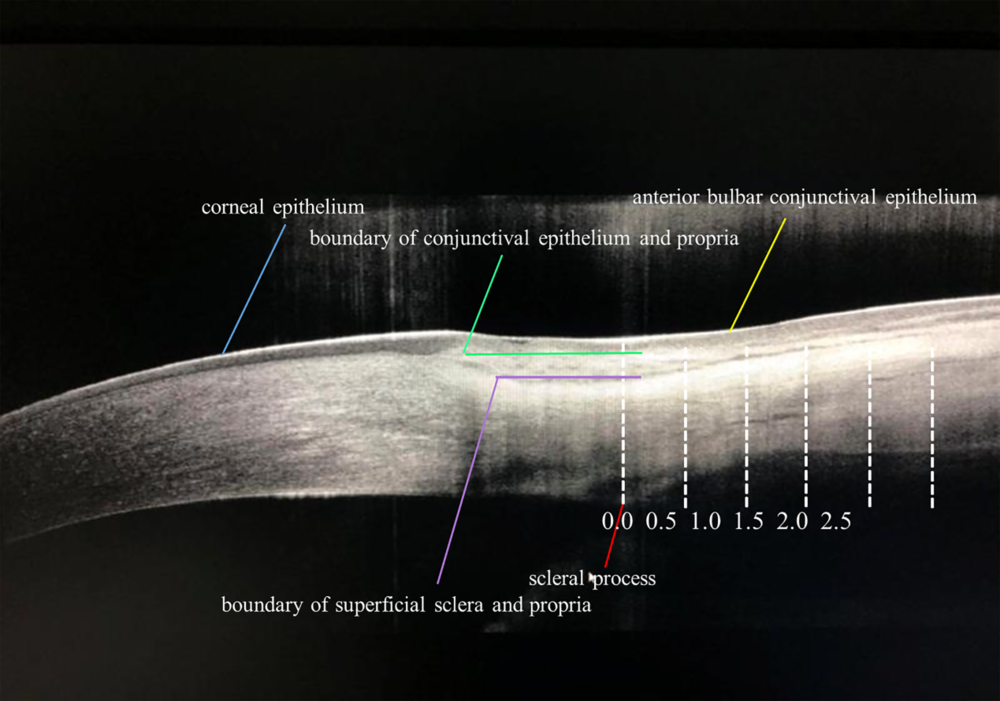 | 图1. 前节OCT图像中参数测量示意图(正常眼)Figure 1. Schematic diagram of a parameter measurement in an AS-OCT image (normal eye). |
病例对照研究。采用SPSS 20.0统计学软件进行数据分析。计量资料采用$\bar{x}\pm s$ 进行描述, 组内各位点比较采用重复测量方差分析, 组间比较采用单因素方差分析, 同时采用LSD-t检验进行两两比较。以P< 0.05为差异有统计学意义。
本研究共纳入25例(30眼), 其中男11例(44%), 女14例(56%); 年龄31~62(51.6± 10.2)岁。其中正常眼组8例(10眼)[(44.8± 6.4)岁], 睑裂斑组8例(10眼)[(56.5± 4.1)岁], 翼状胬肉组9例(10眼)[(56.8± 5.9)岁], 睑裂斑组、翼状胬肉组与正常眼组年龄差异均无统计学意义。所有患者病程1~13(6.0± 3.6)年, 其中睑裂斑组病程1~10(5.3± 3.1)年, 翼状胬肉组病程2~13(6.6± 3.3)年, 2组病程差异无统计学意义。
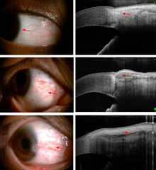
前节OCT的角膜水平横断面图像清晰显示了高反射的睑裂斑与翼状胬肉变性组织贴覆于相应鼻侧角膜、角膜缘和巩膜, 并显示上皮层与其下变性组织结合疏松。在睑裂斑与胬肉组织中, 结膜上皮呈中等反射, 变性组织呈高反射, 浅层巩膜呈低反射, 以上3层不同组织的反射程度不同, 正常结膜上皮下的固有层较均匀无团块, 睑裂斑和翼状胬肉则分别可见暗的固有层团块或上皮下高反射组织团块。见图2。
 | 图2. 正常眼、睑裂斑和翼状胬肉患者的裂隙灯显微镜下外眼像和前节OCT表现 A、C、E:正常眼、睑裂斑和翼状胬肉外眼像; B、D、F:正常眼、睑裂斑和翼状胬肉AS-OCT横断面图像。A、C、E的红色箭头示扫描的区域, B是结膜固有层呈现高反射, D、F是变性组织呈现高反射Figure 2. The appearance of the ocular surface withslit lamp and anterior segment OCT in a normal eyeand in pterygium and pterygium patients. A, C, E: External images of normal, pinguecula and pterygium eyes; B, D, F: Cross-sectional image of normal, pinguecula and pterygium eyes. The red arrows of A, C and E indicate the scanning area. The red arrows of B indicates high reflection of the lamina propria of the conjunctiva. The red arrows of D and F show high reflection of denatured tissues. |
①睑裂斑上皮厚度在各位点与正常眼相比均差异无统计学意义(均P> 0.05); 翼状胬肉在距离巩膜突0.5 mm处较正常眼增厚(均P< 0.05); 翼状胬肉与睑裂斑相比, 结膜上皮层增厚程度在数值上高于后者, 但仅距离巩膜突0.5 mm处差异有统计学意义(P=0.01) (见表1)。②与正常眼相比, 睑裂斑和翼状胬肉均表现结膜固有层增厚(均P< 0.05)。翼状胬肉总体趋势表现为越靠近鼻侧组织增厚越明显, 睑裂斑在各位点均表现增厚(均P< 0.05); 翼状胬肉和睑裂斑相比在距离巩膜突0.0 mm、2.0 mm和2.5 mm位点处增厚程度较高(均P< 0.05), 其余位点差异无统计学意义(见表2)。③与正常眼相比, 睑裂斑和翼状胬肉均表现结膜组织增厚(均P< 0.05)。翼状胬肉和睑裂斑相比在距离巩膜突0.0 mm、2.0 mm、2.5 mm处较厚(均P< 0.05), 其余位点差异无统计学意义(见表3)。
| 表1 正常眼、睑裂斑和翼状胬肉不同位点结膜上皮厚度测量结果(μ m) Table 1 Thickness (μ m) of conjunctival epithelium in normal eyes, pinguecula and pterygium |
| 表2 正常眼、睑裂斑和翼状胬肉不同位点结膜固有层厚度测量结果(μ m) Table 2 Thickness (μ m) of conjunctival propria in normal eyes, pinguecula and pterygium |
| 表3 正常眼、睑裂斑和翼状胬肉结膜总厚度测量结果(μ m) Table 3 Total thickness (μ m) of conjunctiva in normal eyes, pinguecula and pterygium |
在解剖学上, 球结膜由上皮层和其下固有层组成, 其中固有层为上皮下的结缔组织, 含有浅层的腺样层和深层的纤维层[10]。组织病理学研究表明[11], 胬肉组织和睑裂斑组织由表层变性的结膜上皮和其下增殖的胶原纤维和新生血管组成, 是特异性炎症和免疫变态反应导致的弹性组织增生。
球结膜厚度变化的观察对于多种结膜疾病的诊断和治疗评估具有重要意义。既往Feng和Simpson[12]和Zhang等[13]分别利用改装的后节OCT和前节OCT测量正常眼球结膜上皮层、固有层和结膜全层的厚度, 前者报道球结膜上皮厚度为(44.9± 3.4)μ m, 后者则报道球结膜上皮层、固有层和全层厚度分别为(47.3± 8.4)μ m、(190.8± 47.5)μ m和(238.8± 51.1)μ m, 其研究结果与本研究存在一定差异, 分析原因为:①组织形态学上, 球结膜固有层与下方Tenon囊、巩膜的移行没有明显分界, 故其厚度测量本身存在难度[8]; ②本研究中正常眼组的样本量较少, 且年龄限定, 与既往Zhang等[13]的研究(711眼)相比存在量的差异。
活体共聚焦显微镜可用于对两类眼病进行细胞学检测, 但存在花费时间长、范围局限等弊端[11]。本研究首次利用Optovue前节OCT对鼻侧不同位点结膜厚度进行测量, 结果显示:睑裂斑和翼状胬肉均表现为结膜上皮和上皮下结缔组织增厚, 尤以翼状胬肉增厚程度更明显。这可能与翼状胬肉的炎症反应较重有关。此研究方法的建立可定量评估球结膜增生性疾病的病变程度和范围, 为手术方案的设计和治疗结果的随访以及疗效的评估提供较为精准的参数。
本研究在测量中采用间隔0.5 mm, 对鼻侧2.5 mm范围内的6个位点进行测量, 借助巩膜突作为解剖定位可使测量结果更为客观, 具有良好的可重复性与可靠性[14]。但还存在明显的不足, 例如结构图层次的分界是通过人工描画的, 存在一定量的误差, 希望以后信号提取技术的改进能进行自动边界探测; 其次是6个位点的点测量存在定位的误差, 在进一步的研究中, 希望能借助图像处理软件进行6个区域的厚度测量, 进一步提高测量的精确性。
综上所述, 前节OCT由于其非接触性、快捷性、高分辨等特点, 可清晰成像结膜良性增生性疾病眼的结膜组织结构, 可有效进行鉴别诊断, 并可进行较为精准的定量诊断和评估, 为疾病的治疗、随访和疗效评估提供客观的、有实际意义的参数, 其具有广阔的发展前景。
利益冲突申明 本研究无任何利益冲突
作者贡献声明 赵丰平:负责研究设计、收集数据、资料分析及解释、撰写论文, 根据编辑部的修改意见进行修改; 杜持新:负责研究设计、收集数据和质量控制, 指导并修改论文中关键性结果、结论, 并参与编辑部意见修改; 蔡思捷:参与收集数据、参与修改论文并参与编辑部意见修改
The authors have declared that no competing interests exist.
| [1] |
|
| [2] |
|
| [3] |
|
| [4] |
|
| [5] |
|
| [6] |
|
| [7] |
|
| [8] |
|
| [9] |
|
| [10] |
|
| [11] |
|
| [12] |
|
| [13] |
|
| [14] |
|