
OCTA在脉络膜新生血管治疗效果评估中的作用
引用本文
周丽, 吴培培, 徐晶, 陈秀丽, 徐海峰. OCTA在脉络膜新生血管治疗效果评估中的作用[J]. 中华眼视光学与视觉科学杂志, 2019,21(3): 175-180.
Li Zhou, Peipei Wu, Jing Xu, Xiuli Chen, Haifeng Xu. Role of OCTA in Evaluating the Effect of Anti-VEGF Therapy on Choroidal Neovascularization[J]. CHINESE JOURNAL OF OPTOMETRY OPHTHALMOLOGY AND VISUAL SCIENCE, 2019,21(3): 175-180.
Doi: 10.3760/cma.j.issn.1674-845X.2019.03.004Li Zhou, Peipei Wu, Jing Xu, Xiuli Chen, Haifeng Xu. Role of OCTA in Evaluating the Effect of Anti-VEGF Therapy on Choroidal Neovascularization[J]. CHINESE JOURNAL OF OPTOMETRY OPHTHALMOLOGY AND VISUAL SCIENCE, 2019,21(3): 175-180.
Permissions
Copyright©2019, 《中华眼视光学与视觉科学杂志》
《中华眼视光学与视觉科学杂志》 所有
OCTA在脉络膜新生血管治疗效果评估中的作用
第一作者:周丽(ORCID:0000-0002-2812-430X),Email:1154005940@qq.com
摘要
目的: 采用光学相干断层扫描血管成像(OCTA)技术观察抗血管内皮生长因子(VEGF)治疗后脉络膜新生血管(CNV)的形态变化,并探讨OCTA在诊断该类疾病以及在判断预后方面的优势。方法: 回顾性系列病例研究。收集2017年7月至2018年4月在青岛眼科医院就诊的CNV患者17例(17眼)。所有患者常规进行最佳矫正视力(BCVA)、频域相干光断层扫描(SD-OCT)以及OCTA等检查,对患眼行玻璃体腔注射抗VEGF药物(雷珠单抗或康博西普)0.05 ml,治疗后每个月随访1次,采用OCTA观察CNV在治疗前后的变化情况。采用配对 t检验比较治疗前后BCVA和SD-OCT显示的黄斑中心凹视网膜厚度(CMT)的变化。结果: 所有患者治疗前BCVA(LogMAR)为0.69±0.35,治疗后改善到0.49±0.38,差异有统计学意义( t=3.541, P=0.003)。CMT由治疗前的(363±66)μm下降到了治疗后的(302±66)μm,差异有统计学意义( t=4.227, P=0.001)。OCTA显示,抗VEGF治疗后所有患者CNV密度呈不同程度降低;部分患者CNV基本消失,神经上皮层脱离与水肿减轻甚至消失;部分患者虽仍存在粗大的异常血管,但神经上皮层脱离与水肿已完全消失。1例患者虽神经上皮层水肿消失,但仍存在边缘血管吻合,随访3个月复发。结论: OCTA能够清晰显示抗VEGF治疗后CNV的形态变化,可作为临床上CNV的诊断以及治疗效果评估的可靠工具。
关键词:
光学相干断层扫描血管成像; 脉络膜新生血管; 抗血管内皮生长因子
Role of OCTA in Evaluating the Effect of Anti-VEGF Therapy on Choroidal Neovascularization
Abstract
Objective: To observe the morphological changes in choroidal neovascularization (CNV) after anti-vascular endothelial growth factor (VEGF) therapy by using optical coherence tomography angiography (OCTA), and to discuss the advantages of OCTA in diagnosing CNV and judging prognoses.Methods: Seventeen patients (seventeen eyes) diagnosed with CNV in Qingdao Eye Hospital from July 2017 to April 2018 were included in this retrospective case series study. All patients were tested for best corrected visual acuity (BCVA), and regularly examined with spectral domain optical coherence tomography (SD-OCT) and OCTA. All affected eyes were treated with 0.05 ml of intravitreal anti-VEGF agents (Ranibizumab or Conbercept), and all patients were followed up monthly. The changes in BCVA and central macular thickness (CMT) were noted on OCT images and compared with a paired t test. The changes in CNV were observed before and after therapy by using OCTA.Results: The BCVA of (LogMAR) all patients was 0.69±0.35 before treatment, and this indicator was raised to 0.49±0.38 after treatment; the difference was statistically significant ( t=3.541, P=0.003). The CMT of all patients decreased from 363±66 μm before treatment to 302±66 μm after treatment, and the difference was statistically significant ( t=4.227, P=0.001). OCTA showed that the density of CNV decreased by different degrees in all patients after treatment. CNV almost disappeared in some patients, meanwhile, neuroepithelial detachment and edema decreased or completely disappeared. Although OCTA showed the thickened and abnormal vessels in some patients, neuroepithelial detachment and edema disappeared completely on OCTA. While neuroepithelial edema vanished completely, marginal vessel anastomosis could still be seen in one patient, and CNV recurred after 3 months in that patient.Conclusions: OCTA clearly demonstrates the morphological changes in CNV after anti-VEGF therapy, and it can be a reliable tool for the clinical diagnosis and therapeutic evaluation of CNV.
Keyword:
optical coherence tomography angiography; choroidal neovascularization; anti-vascular endothelial growth factor
黄斑部脉络膜新生血管(Choroidal neovascularization, CNV)显著影响视力, 可见于湿性年龄相关性黄斑变性、病理性近视、视网膜血管样条纹、外伤性脉络膜破裂等多种疾病[1], 还有最常见于青年人的找不到明确原因的特发性CNV[2]。由此可见, CNV可发生于各个年龄阶层, 对患者的生活及工作造成严重影响。血管内皮生长因子(Vascular endothelial growth factor, VEGF) 水平升高是CNV发生、发展的重要因素, 因此玻璃体腔注射抗VEGF药物已经成为CNV的一线治疗方法[3, 4]。但抗VEGF药物价格昂贵, 而且有些疾病需要反复治疗。因此, 预测及判断疗效就显得极为重要, 而CNV的类型和面积等指标都是预测和判断抗VEGF药物疗效的重要依据[5, 6]。
目前, CNV的检查方法有荧光素眼底血管造影(FFA)及吲哚菁绿血管造影(Indocyanine green angiography, ICGA), 虽然频域相干光断层扫描(Spectral domain optical coherence tomography, SD-OCT)可以大致判断CNV的类型, 但不能精确显示CNV的构象及面积。近年来, 在SD-OCT基础上发展起来的光学相干断层扫描血管成像(Optical coherence tomography angiography, OCTA)技术, 为CNV的诊断和预后判断提供了一种新的无创性的检查方法。OCTA克服了SD-OCT只能观察病变部位横断面结构的缺点, 能清晰显示CNV的位置、大小和形态等[7]。本研究利用OCTA观察了部分患者在抗VEGF治疗前后CNV的变化情况, 现将结果报告如下。
1 对象与方法
1.1 对象
回顾性分析2017年7月至2018年4月在青岛眼科医院接受抗VEGF治疗的CNV患者的临床资料, 包括一般情况和眼部检查。纳入标准:①经FFA和(或)ICGA确诊黄斑部存在CNV; ②接受过抗VEGF治疗并且随访1次或以上; ③抗VEGF治疗前后均进行过OCTA检查; ④排除同时存在视网膜血管性疾病或炎症等其他眼底病变; ⑤屈光介质透明, 能够保证获得足够清晰的图像。共纳入患者17例(17眼), 其中男14例, 女3例; 年龄29~75(56.6± 14.0)岁。
1.2 检查方法
所有患者均行常规最佳矫正视力(BCVA)检查, 裂隙灯显微镜眼前节检查, 复方托吡卡胺散大瞳孔后眼底检查, FFA或联合ICGA检查, SD-OCT及OCTA检查。BCVA采用国际标准视力表进行检查, 结果转换成最小分辨角对数(LogMAR)视力。SD-OCT检查采用美国Optovue公司的RTVue100-2光学相干断层扫描仪进行, 扫描线长为6 mm, 间隔为30° , 对黄斑区进行放射状线性扫描, 获得经计算机处理产生的视网膜厚度地形图, 同时记录地形图中显示的小凹部位的黄斑中心凹视网膜厚度 (Central macular thickness, CMT)。OCTA扫描采用CIRRUSTM HD-OCT仪(Cirrus HD-OCT 5000, 德国蔡司公司)完成, 黄斑区扫描范围为3 mm× 3 mm, 扫描时先对准患者瞳孔的中心, 然后逐渐推进直至屏幕上出现清晰的眼底图像, 并快速进行拍摄和保存图像。
1.3 玻璃体腔注药
患者取平卧位, 用盐酸丙美卡因表面麻醉成功后, 常规消毒手术视野, 贴无菌手术贴膜并用开睑器开睑。使用5%聚维酮碘溶液冲洗结膜囊。在眼部颞上方角巩膜缘后3.5~4.0 mm处行玻璃体腔穿刺并注射抗VEGF药物(雷珠单抗或康柏西普)0.05 ml, 完成后用0.3%氧氟沙星眼膏或妥布霉素地塞米松眼膏包扎术眼。
1.4 统计学方法
回顾性系列病例研究。采用SPSS 24.0软件进行统计学分析。所有数据符合正态分布, 以均数± 标准差表示。定量资料采用配对t检验进行比较。以P< 0.05为差异有统计学意义。
2 结果
2.1 BCVA和CMT变化情况
抗VEGF治疗后, 患者BCVA(LogMAR)为0.49± 0.38, 较治疗前(0.69± 0.35)有所提高, 差异有统计学意义(t=3.541, P=0.003); 治疗后CMT为(302± 66)μ m, 较治疗前的(363± 66)μ m明显降低, 差异有统计学意义(t=4.227, P=0.001)。
2.2 OCTA图像变化情况
抗VEGF治疗后, 所有患者CNV密度不同程度降低, 血管变得粗大, 边缘吻合支减少; 部分患者CNV基本消失, 相应处B-scan表现为神经上皮层脱离和(或)水肿减轻甚至消失, CNV相对应处反光带变薄或消失(见图l); 部分患者虽仍可见粗大异常血管, 但B-scan神经上皮层的脱离和(或)水肿已完全消失, 即黄斑部呈现“ 干燥” 状态(见图2)。1例患者治疗后血管密度显著降低, 神经上皮层水肿消失, 但CNV边缘血管吻合仍明显, 随访3个月复发(见图3)。
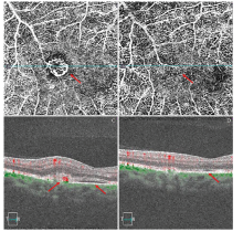
 | 图1. 某例CNV患者抗VEGF治疗前后CNV变化情况 A:治疗前CNV清晰呈线团状(红色箭头所示); B:治疗后CNV基本消失(红色箭头所示); C:治疗前B-scan相应处视网膜下高反射团, 内见血流信号(红色箭头所示), 神经上皮层脱离和水肿(红色箭头所示); D:治疗后B-scan相应处神经上皮层脱离和水肿也基本消失(红色箭头所示)。VEGF, 血管内皮生长因子; CNV, 脉络膜新生血管Figure 1. The changes in CNV before and after anti-VEGF therapy in one CNV patient. A: CNV looks like a ball of strings (red arrow) before therapy. B: CNV almost disappears (red arrow) after treatment. C: High-reflection region and blood flow signal are detected beneath the retina in the corresponding area on OCTA (red arrow), and neuroepithelial detachment and edema (red arrow) are observed before therapy. D: Neuroepithelial detachment and edema disappear completely in the corresponding area on OCTA (red arrow) after treatment. CNV, choroidal neovascularization; VEGF, vascular endothelial growth factor; OCTA, optical coherence tomography angiography. |
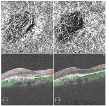
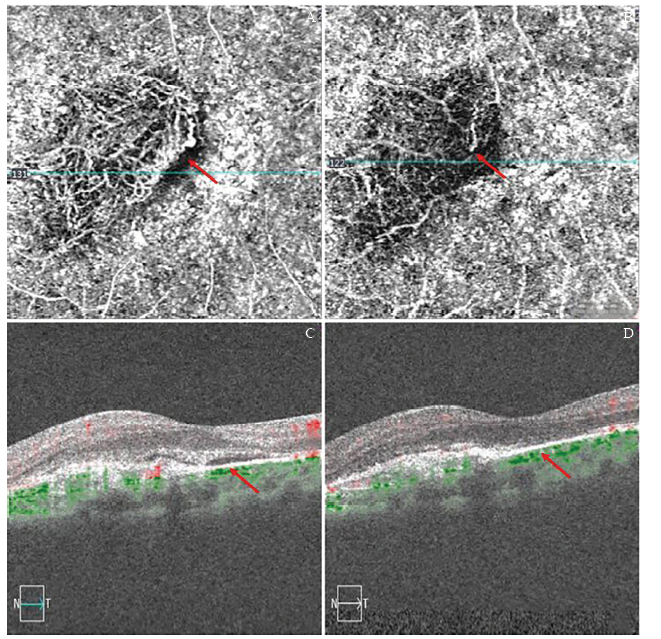 | 图2. 另一例CNV患者抗VEGF治疗前后CNV变化情况 A:治疗前CNV面积较大且血管吻合极为丰富(红色箭头所示); B:治疗后异常血管密度显著降低, 残留粗大血管, 边缘吻合血管基本消失(红色箭头所示); C:治疗前B-scan相应处有异常血流和神经上皮层的脱离与水肿(红色箭头所示); D:治疗后B-scan相应处已无神经上皮层脱离(红色箭头所示), 即黄斑部处于“ 干燥” 状态。VEGF, 血管内皮生长因子; CNV, 脉络膜新生血管Figure 2. The changes in CNV before and after anti-VEGF treatment in another CNV patient. A: CNV is a large area of abundant anastomotic vessels (red arrow) before therapy. B: The density of CNV is obviously reduced and the marginal vessel anastomosis completely disappears, with thickened and abnormal vessels (red arrow) after treatment. C: Abnormal blood flow, neuroepithelial detachment and edema are detected in the corresponding area on OCTA (red arrow) before therapy. D: Neuroepithelial detachment and edema disappear completely in the corresponding area on OCTA (red arrow) after treatment, and the macula is "dry". CNV, choroidal neovascularization; VEGF, vascular endothelial growth factor; OCTA, optical coherence tomography angiography. |
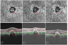
 | 图3. 随访复发的CNV患者抗VEGF治疗前后CNV变化情况 A:治疗前CNV清晰明显(红色箭头所示); B:治疗后2个月CNV密度显著降低, 但仍存在明显的边缘血管吻合(红色箭头所示); C:治疗后随访3个月CNV复发, 血管密度较前增高(红色箭头所示); D:治疗前B-scan相应处有丰富血流信号及神经上皮层脱离与水肿(红色箭头所示); E:治疗后2个月B-scan相应处神经上皮层水肿消失(红色箭头所示); F:治疗后随访3个月CNV复发, B-scan相应处有明显的神经上皮层脱离(红色箭头所示)Figure 3. The changes in CNV before and after anti-VEGF treatment in one recurrent CNV patient. A: CNV is clear and obvious (red arrow) before treatment. B: The density of CNV is obviously reduced, but the marginal vessel anastomosis is still obvious (red arrow) after 2 months of treatment. C: CNV recurs after 3 months and the density of CNV is high (red arrow). D: Rich blood flow signal, neuroepithelial detachment and edema are detected in the corresponding area on OCTA (red arrow) before treatment. E: Neuroepithelial edema disappears in the corresponding area on OCTA (red arrow) after 2 months of treatment. F: Neuroepithelial detachment recurs in the corresponding area on OCTA (red arrow) after 3 months. CNV, choroidal neovascularization; VEGF, vascular endothelial growth factor; OCTA, optical coherence tomography angiography. |
3 讨论
近年来, OCTA作为眼科影像学的一项革命性突破已经广泛应用于临床, 因其无创和操作便捷等特点[8], 不但可以部分替代FFA, 而且在某些情况下比FFA更具优越性而广泛应用于眼底疾病的诊断和发病机制的研究, 在CNV的诊断以及抗VEGF治疗后疗效的评估中也起着重要作用[9]。有研究表明, 对于CNV的诊断结果, OCTA与FFA呈现出良好的对应关系[10], 由此也可以说明OCTA对于CNV的诊断可靠性极强。通过OCTA显示的血管成像图片与SD-OCT呈现的相应横断面结构信息相结合, 可以客观评估CNV的动态变化[11], 并为后续治疗提供可靠依据。本研究中, 治疗前所有患者均可通过OCTA清晰显示CNV的大小和形态, 经典型CNV与FFA造影所显示的形态有很好的一致性; 治疗后OCTA图像中可以清晰显示脉络膜血管密度降低, 这种直观对比可以为判断患者抗VEGF应答情况提供可靠依据。CNV中细小血管及边缘的血管吻合往往提示CNV的活动性[12, 13], 在本研究中有一位患者经抗VEGF治疗后SD-OCT显示神经上皮水肿消失, OCTA显示细小血管明显消失, 但边缘血管吻合仍然存在, 停止治疗后3个月复发。因此, 提示治疗过程中需要高度警惕OCTA中提示CNV活动性的征象, 从而适时对治疗方案做出调整, 为不同的患者提供个性化治疗, 以达到改善患者远期预后的目的。
与传统的CNV检查方法FFA相比, OCTA有其独特的优势:不需要造影剂, 从而避免了药物过敏的风险[14]; 没有造影剂渗漏, 可以清晰显示血管的结构, 但伴随而来的是不能动态观察血流情况, 从而不能判断CNV是否仍然具有高通透性(CNV活动性指标), 此时需要结合B-scan显示的断层结构正确判断治疗时机。在研究中, 部分患者抗VEGF治疗后虽然OCTA明确显示仍然存在异常血管, 但B-scan相应处的神经上皮层已无水肿, 说明CNV虽然存在, 但管壁通透性已不大, 异常血管趋于“ 成熟” 。如果此时继续注射价格昂贵的抗渗漏因子(抗VEGF药物), 追求异常血管完全消失则得不偿失。此外, OCTA的出现使得许多“ 亚临床” CNV被发现, 但对此类病变治疗与否尚无定论[15, 16]。
目前, 尽管OCTA在临床应用中优势显著, 但仍在检查方面存在一定的局限性。例如, 对患者的配合要求相对较高, 患者需要较长时间注视, 否则会影响图像质量; 血流速度过快或过慢的异常血管都不能被检测到, 因此不能显示所有的CNV, 所以即使OCTA未检测到CNV, 也不能排除CNV存在的可能性。虽然本组患者纳入的全部是治疗前能够清晰显示CNV的患者, 但与FFA相比, 部分患者OCTA图像中的异常血管较FFA中显示的明显较少, 这是由OCTA成像原理所造成的[17, 18]。因此, 期待未来OCTA在发展和改进的过程中能克服上述缺陷, 为CNV提供更为精确和可靠的检查。
综上所述, OCTA在诊断CNV以及在判断预后中发挥着至关重要的作用, 但因本研究样本含量较少, 而且没有量化分析CNV的血管形态和直径等指标的改变情况, 因此仍存在一定的不足之处。在未来的研究过程中还需要扩大样本含量, 进一步采用定量的方法分析CNV在抗VEGF治疗前后各个指标的改变情况, 以此更加深入地了解OCTA在诊治CNV方面的应用, 为临床实践提供更加可靠的指导。
利益冲突申明 本研究无任何利益冲突
作者贡献声明 周丽:收集数据; 资料的分析和解释; 撰写论文; 根据编辑部的修改意见进行修改。吴培培:收集数据; 参与选题。徐晶:收集数据; 参与选题。陈秀丽:收集数据; 参与选题。徐海峰:收集数据; 参与选题、设计、资料的分析和解释; 修改论文中关键性结果、结论; 根据编辑部的修改意见进行核修
The authors have declared that no competing interests exist.
参考文献
| [1] |
|
| [2] |
|
| [3] |
|
| [4] |
|
| [5] |
|
| [6] |
|
| [7] |
|
| [8] |
|
| [9] |
|
| [10] |
|
| [11] |
|
| [12] |
|
| [13] |
|
| [14] |
|
| [15] |
|
| [16] |
|
| [17] |
|
| [18] |
|